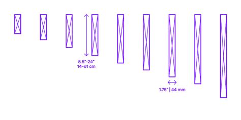
lv thickness | lvl dimensions chart lv thickness Increased left ventricular myocardial thickness (LVMT) is a feature of several cardiac diseases. The purpose of this study was to establish standard reference values of normal LVMT with cardiac magnetic resonance and to assess variation with image acquisition plane, demographics, and left ventricular function. Find ACDelco DOT 4 GMW Brake and Clutch Fluid and get Free Shipping on Orders Over $109 at Summit Racing! ACDelco DOT4 GMW fluid features the same glycol base as DOT 3 fluid but handles more heat. As a superior brake and hydraulic clutch fluid for systems requiring performance level fluid, ACDelco DOT 4 GMW helps preserve internal .
0 · lvl standard sizes chart
1 · lvl dimensions chart
2 · lvl catalogue
3 · left ventricular hypertrophy symptoms
4 · left ventricular hypertrophy risk
5 · left ventricular hypertrophy causes
6 · Lv thickness echo
7 · 4x12 lvl beam dimensions chart
Currency Converter. US-Dollar - Lew. United States dollar to Bulgarian lev (USD to BGN) Quickly and easily calculate foreign exchange rates with this free currency converter. From. Switch..
Our LV calculator allows you to painlessly evaluate the left ventricular mass, left ventricular mass index (LVMI for the heart), and the relative wall thickness (RWT). Read on and discover all the details of our LV mass .
Left ventricular hypertrophy changes the structure of the heart and how the heart works. The thickened left ventricle becomes weak and stiff. This prevents the lower left heart chamber from filling properly with blood. Our LV calculator allows you to painlessly evaluate the left ventricular mass, left ventricular mass index (LVMI for the heart), and the relative wall thickness (RWT). Read on and discover all the details of our LV mass calculator and its variables: Definitions of abnormal LV mass index; PWd normal range; and; IVSd in echo ️ Increased left ventricular myocardial thickness (LVMT) is a feature of several cardiac diseases. The purpose of this study was to establish standard reference values of normal LVMT with cardiac magnetic resonance and to assess variation with image acquisition plane, demographics, and left ventricular function.
Normal Values - Echopedia. Below an up-to-date list of echocardiographic normal values. Contents. 1 Left Ventricle. 1.1 Left Ventricular Systolic Function. 1.2 Left Ventricular Diastolic Function. 1.3 Left Ventricular Mass and Geometry. 1.4 Left Ventricular Size. 2 Right Ventricle. 2.1 Right Ventricular and Pulmonary Artery Size.Normal values for LV chamber dimensions (linear), volumes and ejection fraction vary by gender. A normal ejection fraction is 53-73% (52-72% for men, 54-74% for women). Refer to Table 2 (normal values for non-contrast images) and Table 4 (recommendations for the normal Left ventricular hypertrophy is thickening of the walls of the left ventricle, the heart’s main chamber. The left ventricle pumps blood into the aorta (the largest artery in the body), which sends this oxygenated blood to tissues throughout your body. Normal sex- and age-specific reference ranges for left ventricular mid-diastolic wall thickness (LV-MDWT) at prospective electrocardiographically triggered mid-diastolic CT angiography were provided, and LV-MDWT was strongly correlated with .
Normal sex- and age-specific reference ranges for left ventricular mid-diastolic wall thickness (LV-MDWT) at prospective electro-cardiographically triggered mid-diastolic CT angiography studies were provided. The upper limit of LV-MDWT for any . Left ventricular mass, wall thickness, and the ratio of wall thickness to radius are important measures to characterize the spectrum of left ventricular geometry. For clinicians, an increase in left ventricular mass is the hallmark of left ventricular hypertrophy. Left ventricular hypertrophy, or LVH, is a term for a heart’s left pumping chamber that has thickened and may not be pumping efficiently. Sometimes problems such as aortic stenosis or high blood pressure overwork the heart muscle.
Left ventricular hypertrophy changes the structure of the heart and how the heart works. The thickened left ventricle becomes weak and stiff. This prevents the lower left heart chamber from filling properly with blood. Our LV calculator allows you to painlessly evaluate the left ventricular mass, left ventricular mass index (LVMI for the heart), and the relative wall thickness (RWT). Read on and discover all the details of our LV mass calculator and its variables: Definitions of abnormal LV mass index; PWd normal range; and; IVSd in echo ️ Increased left ventricular myocardial thickness (LVMT) is a feature of several cardiac diseases. The purpose of this study was to establish standard reference values of normal LVMT with cardiac magnetic resonance and to assess variation with image acquisition plane, demographics, and left ventricular function. Normal Values - Echopedia. Below an up-to-date list of echocardiographic normal values. Contents. 1 Left Ventricle. 1.1 Left Ventricular Systolic Function. 1.2 Left Ventricular Diastolic Function. 1.3 Left Ventricular Mass and Geometry. 1.4 Left Ventricular Size. 2 Right Ventricle. 2.1 Right Ventricular and Pulmonary Artery Size.
Normal values for LV chamber dimensions (linear), volumes and ejection fraction vary by gender. A normal ejection fraction is 53-73% (52-72% for men, 54-74% for women). Refer to Table 2 (normal values for non-contrast images) and Table 4 (recommendations for the normal
Left ventricular hypertrophy is thickening of the walls of the left ventricle, the heart’s main chamber. The left ventricle pumps blood into the aorta (the largest artery in the body), which sends this oxygenated blood to tissues throughout your body. Normal sex- and age-specific reference ranges for left ventricular mid-diastolic wall thickness (LV-MDWT) at prospective electrocardiographically triggered mid-diastolic CT angiography were provided, and LV-MDWT was strongly correlated with .Normal sex- and age-specific reference ranges for left ventricular mid-diastolic wall thickness (LV-MDWT) at prospective electro-cardiographically triggered mid-diastolic CT angiography studies were provided. The upper limit of LV-MDWT for any .
lvl standard sizes chart
Left ventricular mass, wall thickness, and the ratio of wall thickness to radius are important measures to characterize the spectrum of left ventricular geometry. For clinicians, an increase in left ventricular mass is the hallmark of left ventricular hypertrophy.
lvl dimensions chart
burberry pomegranat pink lipstick

burberry lipstick limited edition

lvl catalogue
Dr. Dongsi Lu is a pathologist in Chesterfield, MO and is affiliated with St. Luke's Hospital. She received her medical degree from Beijing Medical University and has been in practice 16 years. She specializes in dermatopathology, anatomic pathology, and clinical pathology.
lv thickness|lvl dimensions chart


